Showing posts with label Microbe Monday. Show all posts

Monday, October 25, 2010

Microbe Monday: O/OREOS, Microbes in Space

Microbes are going to orbit the Earth's atmosphere for six months, the first mission since the 1970s to test how a life form handles the conditions outside the protection of the earth's magnetic field, according to Space Daily.

The O/OREOS (Organism/Organic Exposure to Orbital Stresses) astrobiology nanosatellite mission goal is to demonstrate the capability to do low-cost science experiments on autonomous nanosatellites in space in support of the Astrobiology Small Payloads program. Although the NASA Ames Small Spacecraft division manages the O/OREOS mission, all of the operations are conducted by staff and students from the Robotic Systems Laboratory at Santa Clara University. The mission should help answer Astrobiology's fundamental questions on the origin, evolution and distribution of life. (How's that for a kick-ass dissertation!?!)

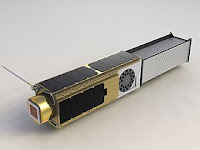
In an effort to assess survival and adaptation to stress, the microorganisms Halorubrum chaoviatoris and Bacillus subtilis will orbit the Earth in a 5.5kg automated laboratory. The spacecraft, which is about the size of a loaf of bread, is equipped with sensors that will monitor the internal pressure, temperature, humidity, radiation and acceleration while communications system regularly transmit data back to earth for scientific analysis. The organisms will reside within a self-contained vessel that is regulated for pressure, humidity and temperature. Growth media will be provided to the organisms as they are exposed to the high-energy radiation and microgravity conditions. After O/OREOS reaches orbit, the experiment will initiate and begin to rehydrate and expose three sets of the microbes to media at three different times: a week, three months and six months after launch. A three-color LED and detector will monitor growth and metabolism using a colorimetric assay.

A second experiment on the coolest space-lab evah will monitor the stability and changes in four classes of organic matter as they are exposed to space conditions. The stability will be assessed by studying in-situ changes in UV, visible and near-infrared light absorption through a daily measurement.


Monday, October 18, 2010

Microbe Monday: 1/2 lesson, 1/2 rant

Last week at the International Conference on Gram-positive Pathogens, there were talks (and posters) on Bacillus anthracis and Clostridium difficile. While listening to some of these talks and poster presentations, I was forced to endure a phrase that grinds my nerves. What is that you ask? It's the phrase: "The dormant spore."


It bugs me because it's (1) incorrect and (2) redundant.

(1) Why the phrase is incorrect.

Bacteria don't make spores. Members of the genera Bacillus and Clostridium undergo sporulation, but the product is an endospore, not a spore. A spore is the result of asexual reproduction, an endospore is not.

Now, I realize that if you are giving a talk about endospore or sporulation that you are going to say the word endospore multiple times. I don't think it is a big deal if people drop the word endospore for spore for the duration of the talk if they at least refer to it correctly in the beginning. This is fairly common. Referring to an endospore as a spore didn't even use to bother me at all until I met multiple people working on endospore-forming bacteria that did not realize the difference.

(2) Why it's redundant.

An endospore is a dehydrated, highly-refractile product resulting from a series of biochemical and morphological changes known as sporulation. Endospores are resistant to ultra-violet radiation, extreme temperatures, chemical disinfectants, dessication and pressure. They are metabolically inactive, i.e. dormant.

Using the phrase dormant spore (or dormant endospore) is the same as saying frozen ice.

It might seem silly, but it bugs me.

Anyone want to share their scientific pet peeves?

Sunday, August 29, 2010

Microbe Monday: A sterile stick and other tools

My previous advisor used to joke that all you need to do microbiology is a sterile wooden stick. You can use this simple tool too isolate colonies, pick colonies, inoculate media and isolate DNA*. If you were to visit my bench, you would find a bucket of sterile sticks as I use them almost every day. However, I need a lot more than a wooden stick to get my research done. The machines I use most often are the thermocycler, FPLC, biacore and any kind of spectrophotometer. Unfortunately, my favorite tool, the microscope, is one I rarely use.
One of the major reasons I love this instrument because it enables me to actually see the microbes I am studying. Since my work typically involves using RNA, DNA or protein, I spend most of my time looking at tubes containing a clear solution, or a if I'm lucky, a solution containing a dye. If not a tube, then I'm probably viewing a stained gel (acrylamide or agarose). It's just not as visually stimulating as looking at something under the microscope.**
Another reason I like to use the microscope is because you can take pictures or video of what you observe. Pictures and videos typically go over really well in a seminar and when applicable, they are great in a paper. The presence of images or video in a paper was often a deciding factor when I was deciding on a paper for journal club. I think everyone agrees that it's nice to see something besides a graph or table every now and then.***
For researchers like me, there just isn't usually a reason to have photos or video from a microscope. However, when I can, I choose assays that utilize a microscope. For example, to assess translation, I'll use a reporter gene like GFP so that not only do I get to use the microscope, but I get to look at fluorescence and that is way cool. I've also used fluorescence to assess protein-protein interactions. In both of these cases I can use the microscope for qualitative "yes or no"-type of data and then if need be, I can use a fluorometer for more quantitative data.
To illustrate my point, here are images from wikimedia commons. I wanted to include videos, but blogger wasn't cooperating.****


Original figure legend: Multiple fluorescence 2PE imaging. 2PE multiple fluorescence image from a 16 μm cryostat section of mouse intestine stained with a combination of fluorescent stains (F-24631, Molecular Probes). Alexa Fluor 350 wheat germ agglutinin, a blue-fluorescent lectin, was used to stain the mucus of goblet cells. The filamentous actin prevalent in the brush border was stained with red-fluorescent Alexa Flu or 568 phalloidin. Finally, the nuclei were stained with SYTOX ® Green nucleic acid stain (2).


Polymicrobic biofilm grown on a stainless steel surface in a laboratory potable water biofilm reactor for 14 days, then stained with 4,6-diamidino-2-phenylindole (DAPI) and examined by epifluorescence microscopy (3).


*Once the DNA is precipitated, you can wind it around a sterile stick or toothpick in leu of centrifugation. This is not really effective if you aren't isolating a large quantity of DNA.
** Yes, you can visualize your data in ways that don't involve a microscope and as a general rule it's awesome to obtain data, be that data in the form of a band, a sensogram or absorbance reading, but in my opinion seeing your data in a living organism is way cool.
***Not that anything is wrong with a table or graph. Pretty much all my data is presented in this form.
****I know that there are many compelling images and videos utilizing fluorescence microscopy, but I am not sure what the rules are regarding image and video usage from scientific papers. If anyone would like to enlighten me, please feel free. If anyone knows of some better images, that I can post without committing any kind of offense or spending money, please let me know and I will be happy to post them.


(2) Multi-photon excitation microscopy. BioMedical Engineering OnLine, 2006, 5:36.DOI:10.1186/1475-925X-5-36.
(3) Centers for Disease Control and Prevention Rodney M. Donlan: "Biofilms: Microbial Life on Surfaces"

Sunday, April 4, 2010

Microbe Monday: Awesomely-strong glue

Its shape, adhesive properties, intricate cell-cycle regulation and asymmetric cell division are a just few of the reasons why Caulobacter crescentus is a well-studied microorganism (1). In nature, C. crescentus is typically found attached to submerged surfaces within aquatic environments via a stalk structure which protrudes from a single pole of the cell. An adhesive holdfast, located at the tip of the stalk (Fig. 1), composed of N-acetylglucosamine (sugar molecules) (Fig. 2) and other unknown substances exhibits the strongest adhesion force of any known natural material (2, 3, 4).
The majority of the C. crescentus life cycle is spent in the adherent stalked form. However, the early third of the life cycle is spent as a motile, swarmer cell (Fig. 1). It is hypothesized that the swarmer cells, which exhibit a single polar flagella, can explore new environments where nutrients are more plentiful. Once the flagella is shed, a stalk and holdfast are synthesized in the same location and promote adherence to a surface. Stalked cells undergo cell division and produce new swarmer cells, which begin the process anew (Fig. 1)(1, 5).


Figure 1. C. crescentus undergoing cell division. The stalked cell is located on the top, with the stalk and holdfast at the top of the frame. The swarmer cell is located underneath such that the flagella is protruding toward the bottom right-hand corner of the image. (This image was obtained from MicrobeWiki courtesy of Yves Brun.)

The glue: What is it and how strong is it?
In addition to other unknown elements, the holdfast is composed of oligomers of N-acetylglucosamine (Fig. 2). Treatment of the holdfast with lysozyme, an enzyme that will cleave N-acetylglucosamine oligomers, reduces the adhesive force to less than 10% (3, 4). Furthermore, C. crescentus mutants lacking the sugar molecules at the tip exhibit a significant adherence defect. It was determined that the force required to remove C. crescentus from a glass surface is over 70 Newtons per square millimeter or 5 tons per square inch (2). This force is equivalent to the downward force exerted by three cars balancing on a quarter (6).

Figure 2. N-acetylglucosamine monomer (a.k.a GlcNac, NAG).

That's pretty cool, but why should anyone care?
Unlike superglue, the adhesive substance produced by C. crescentus is non-toxic and it adheres well under water, both the fresh and salt varieties. Potential applications include, biodegradable surgical and dental adhesives and repair of surfaces exposed to water (2, 6). Furthermore, studying adherence of C. crescentus should provide insight into biofouling and biofilm formation (2).

Why so sticky?
It is thought that in its natural environment, C. crescentus attached near the surface of water must contend with the passage of waves at the air-liquid interface, which exerts a significant force (2).

References:
(1) Brown et al. Complex regulatory pathways coordinate cell cycle progression and development in Caulobacter crescentus. Adv Microb Physiol. 2009; 54:1-101.

(2) Tsang et al Adhesion of Single Bacterial Cells in the Micronewton Range. PNAS. 2006; 103(15):5764-5768.

(3) Smith et al Identification of Genes Required For Synthesis of the Adhesive Holdfast in Caulobacter crescentus. J. Bacteriol. 2003; 185:1432-1442.

(4) Li et al. The Elastic Properties of the Caulobacter crescentus Adhesive Holdfast are dependent on Oligomers of N-Acetylglucosamine. J. Bacteriol. 2005; 187(1):257-265.

(5) Jenal. The Role of Proteolysis in the Caulobacter crescentus Cell Cycle and Development. Research in Microbiology 2009; 160:687-695.

(6) Iddo Genuth & Lucille Fresco-Cohen. Nature's Superglue The Future of Things (2006)

Monday, March 22, 2010

Microbe Monday: 10 Favorite Microbes

A while ago, fellow microbiologist and blogger, Thomas Joseph, started a meme of sorts on his blog asking others to list their 10 favorite microbes. Quite a few bloggers put a list together, and I intended too, but for one reason or another, I never did.
Well, I'm doing it now and using it for my latest Microbe Monday installation. Over the next several weeks I will use Microbe Monday to fully geek out about why each of these organisms made the list.* For this post, I'll keep my inner-nerd contained and just give you a little hint as to why they made the list.

1. Caulobacter crescentus: strong-ass natural glue, wicked-cool asymmetrical cell division.
2. Listeria monocytogenes: utilizes an "actin rocket" for one of the most unique methods of spreading from one host cell to another. Just wait until you see the movie.**
3. Dictyostelium discoideum: Individual amoebas can work together and move as a slug. A freaking slug. It leaves a trail and everything.
4. The Bacillus cereus group species: Almost exactly the same...almost.
5. Streptomyces: Produce over half of the world's clinically useful, naturally-derived antibiotics.
6. Deinococcus radiodurans: It's the world's toughest bacterium according to The Guinness Book of World Records.
7. Bacillus subtilis: microbial cannibalism at its finest.
8. Staphylococcus aureus: Amazing ability to evade the host's immune system and strike fear into the hearts of ordinary citizens, forcing them to learn acronyms like MRSA and maybe even understand the concept of antibiotic resistance.
9. Vibrio fischeri: Quarum sensing, bioluminescence, what's not to love?
10. Escherichia coli: Come on. I don't need to explain this one.

*will not be necessarily be back-to-back.

** provided I can find one that I won't get in trouble for posting.

Monday, March 8, 2010

Microbe Monday: Way cooler than an ant farm.

As a child, I did not own an ant farm. I thought they were pretty cool, but there was something about keeping a colony of ants in the house that just didn't sit to well with my mother. I did own Sea Monkeys, but I accidentally boiled them to death in the window.* Anyway, I think I found something even more cool than an ant farm or sea monkeys. While perusing the ASM website, I decided to check out the 2009 Editor's Choice Award Winners, where I found the video: "Mud and Microbes: A Time-lapse Photographic Exploration of a Sediment Bacterial Community."
Using a combination of time-lapse photography, light, an adaptation of a Winogradsky column, sediment from a pond, finely-shredded paper towels, calcium carbonate and magnesium sulfate, the participants created a simple, yet fascinating way to get a peek at microorganisms in the soil and sediment. The creators of the following video hope that it will serve as a catalyst to discuss microbial ecology and microorganism dynamics in the world around us and to increase increase interest in the study of soil microorganisms in nutrient cycling.
The video is composed of stills taken over the 40 day experiment and the progression clearly demonstrates how phototropic microbes with differing metabolic capabilities respond to the nutrients and light.


In a longer version of the video the narrator informs us that the bottom area of the plates become anaerobic, favoring reducing conditions where sulfate-reducing and cellulose-degrading bacteria proliferate. The black color is iron sulfide. (Note: the shredded paper towels are located in the bottom of the "mud column.") He goes on to describe that the pink/purple areas near the bottom are likely populated by purple, nonsulfur proteobacteria, the green patches likely represent green and purple sulfur proteobacteria, while the green at the very top is most likely green algae and/or cyanobacteria. It is important to note that the bacteria from this experiment were not isolated or typed and these descriptions are just best guesses.

Anyway. I kinda want to make one of these, take samples and look at them under the scope.

*Portions of the Sea Monkey "aquarium" contained magnifying glass that would enlarge the Sea Monkey as it swam by, allowing one to see that the tiny things swimming around in that container didn't actually look like monkeys. Sun, water and magnifying glass is apparently a deadly combination for a Sea Monkey.


Authors
Michael Lemke
Microbial Ecology
University of Illinois at Springfield
Springfield, IL 62701
USA
Email: mlemk1@uis.edu

Roza George
Department of Microbiology
University of Georgia

Keith Miller
Department of Computer Science
University of Illinois at Springfield
Springfield, IL 62703-507

References:
1. Charlton, P. J., J. E. McGrath, and C. G. Harfoot. 1997. The Winogradsky plate, a convenient and efficient method for enrichment of anoxygenic phototrophic bacteria. J. Microbiol. Methods 30:161–163.
2. Couger, G. 2002. Habitat for lab specimens and other uses for common household items.http://www.microscopy-uk.org.uk/mag/artaug02/gchabitat.htm.
3. Rogan, B., M. Lemke, M. Levandowsky, and T. Gorrell. 2005. Exploring the sulfur nutrient cycle using the Winogradsky column. Am. Biol. Teacher67:279–287.
Music.
Barbara Schubert and theUniversity of Chicago Orchestra performed Richard Straus’s Also Sprach Zarathurstra,http://www.archive.org/details/uso20000527. Creative Commons license: attribution, noncommercial, no derivative works.